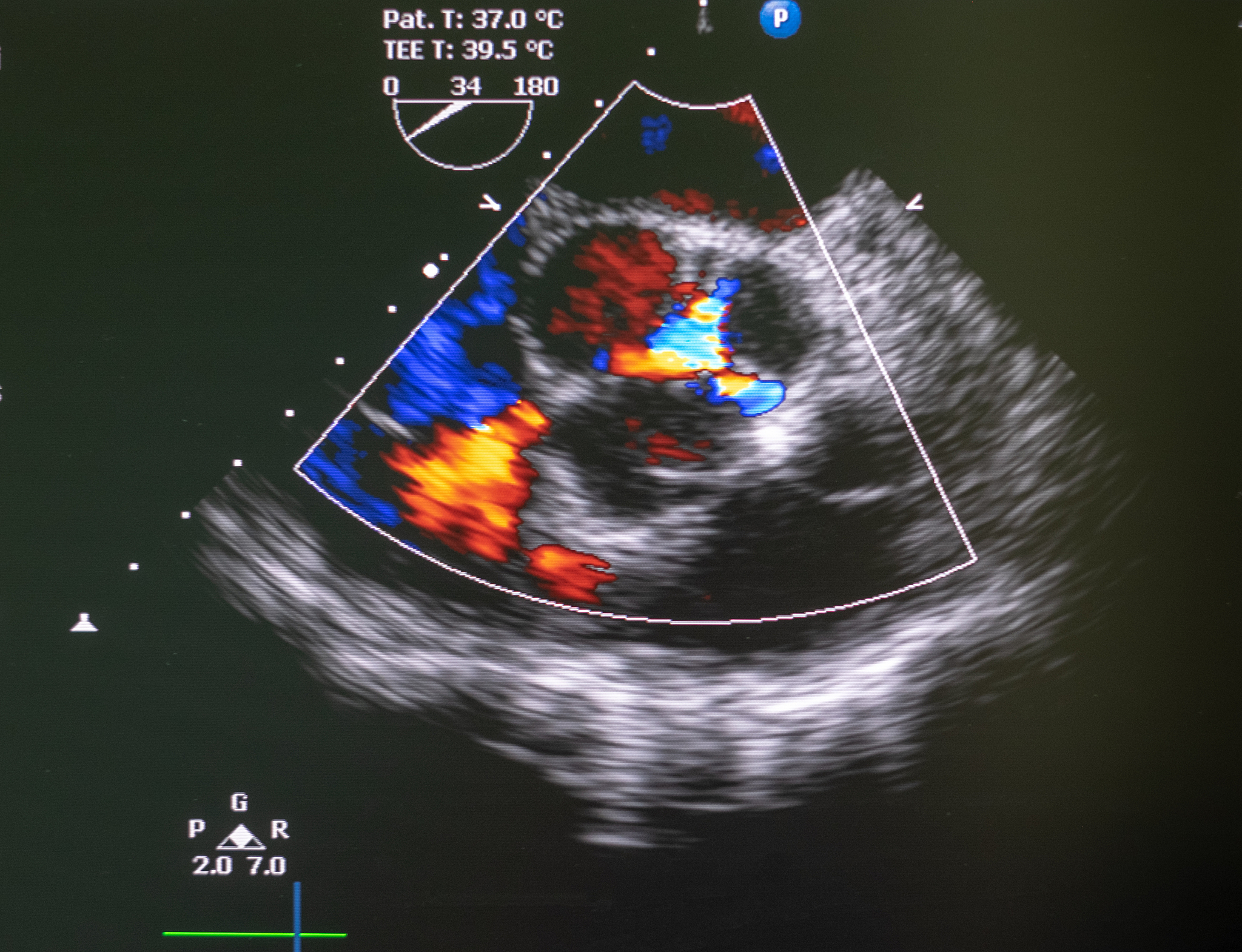
右侧颈总动脉分叉处斑块致颈内动脉狭窄 - 超声检查结果
Routine ultrasound examination: A slightly 'hypoechoic' plaque is observed at the bifurcation of the right common carotid artery to the beginning of the internal carotid artery, leading to stenosis of the starting segment of the internal carotid artery. The maximum flow velocity at the stenosis is about 272cm/s, and the flow velocity near the proximal end of the stenosis is about 81cm/s. The PSVICA/PSVCCA is 3.36. According to the NASCET standard, the diameter stenosis rate is approximately 58.4%. A low 'hypoechoic' attachment is faintly visible at the distal end of the plaque, which shows slight movement with the pulsation of the carotid artery. The color Doppler also shows a suspicious filling defect.

原文地址: https://www.cveoy.top/t/topic/fZbI 著作权归作者所有。请勿转载和采集!